

FEMORAL
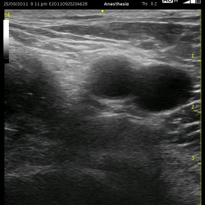
The femoral nerve is seen to the left of the femoral artery and vein as a hyperechoic (white) triangle in the ultrasound image to the left. This is often one of the first nerve block to master with ultrasound because it is a relatively simple and superficial option and also probably because Total Knee Arthroplasties are such common and such painful procedures. The sonoanatomy is relatively straight-forward, however, as I will point out, there are a number of nuances to this procedure that should be kept in mind in order to avoid some minor and some serious errors.
In the cadaver image below, the femoral nerve is seen proximally as a solid and flat structure that fans out significantly as it travels distally. Note the sartorius muscle laterally. You will be looking for this landmark during this nerve block. It is difficult to appreciate the fascia iliaca which thinly covers the femoral nerve and the iliopsoas muscle. I have made the comment that recognizing this structure on ultrasound is more important to performing this nerve block than identifying the nerve, and I will elaborate further on this later. Utilizing an entry as lateral as the sartorius muscle (and often through it), I view the needle with ultrasound passing in a relatively flat plane to the transducer. Be sure to identify the femoral nerve as proximally as possible, and then to find a good entry point in the skin, slide your probe laterally instead of along the inguinal crease. This is an easy mistake to make, and I will discuss later why this is important. Determine the appropriate trajectory toward your target, and follow your needle as you move it medially. Before reaching the femoral nerve, [wlm_ismember]I dive slightly deeper toward the iliopsoas muscle feeling for the ‘pop’ as I cross the fascia iliaca which is tightly bound to the muscle. Remember, you can ‘pop’ through only the fascia lata here, and your block will be unsuccessful. Once under the fascia, I inject a few ml’s of local to confirm that I am actually under it and to create a ‘track’ for my needle to follow as I advance toward the nerve. This is confirmed by seeing a thin but distinct black line appearing under the fascia iliaca that will not track over the femoral vessels. You should not see ‘muscle stranding’ (indicating that you pushed too deep and into the iliopsoas muscle. Just pull back and reinject to find the effect you are after if this happens. At this point, I advance my needle toward the nerve as I add a few more ml’s of local and watch for the nerve to become more easily visible (‘enhancement’) which should occur due to the local being in proximity to it. I will attempt to direct the needle under the femoral nerve and continue injecting as I watch for circumferential spread around it. I leave the catheter under the nerve on the medial aspect unless it takes too long to accomplish or the local does not provide a path that I can take without serious manipulation of the nerve. You MUST get under the fascia iliaca, but just TRY to get under the nerve. This will theoretically bathe a little more the posterior division which is responsible for the more painful sources like the joint, femur and muscle. If you ever see local spreading over the femoral vessels, you know you are not in the correct position with the injection, and you must readjust your needle to get below the fascia iliaca.[/wlm_ismember]

[nonmember]…
REGISTER for FREE to become a SUBSCRIBER or LOGIN HERE to see the full article!
[/nonmember]
[wlm_ismember]
The needle is in the final position in this picture where the majority of the injection takes place. The catheter is advanced a centimeter or so beyond this point. Again, note the relatively flat plane the needle is held in so that the ultrasound is easily able to visualize it (including the hallmark curved bevel at the end of the Tuohy). The skin entry should be somewhere near the last two black lines at the bottom of the picture over the sartorius muscle so that the catheter stays in place and to minimize ‘track back’ issues. ‘Track back’ is the return of local anesthetic outside the catheter toward the skin. Once local exits the skin, the sticky products will no longer be sticky, and tegaderms become wet and begin to loosen. Extending the distance that the catheter is beneath the skin and traversing tissues like muscle and the fascia iliaca will minimize the impact of ‘track back’. As mentioned above, if you enter near the inguinal crease, your tegaderm will loosen at the edges with hip flexion, causing it to loosen prematurely and dry out your adhesives below. This will lead to leaking catheters, and this is never acceptable.

If you select a path toward the femoral nerve that is too distal, you can see that a few things happen. First, the femoral nerve becomes less distinct. More importantly, the femoral vein begins to position itself deep to the nerve, and the femoral artery splits off the deep femoral artery. If you lose the end of your needle more proximally, no big deal. If you do so more distally, you are likely to impale one of these three vessels. Take a look at the Archetypal Images below which demonstrate the sonoanatomy at a proximal and at a more distal location. In addition to the vascular changes that occur, note how the sartorius muscle slides toward the femoral nerve and vessels in the more distal location (The movement of the sartorius muscle is also key in locating the best place to perform the fascia iliaca block.). These are all clues that you can use to help you find the best place to enter the skin and perform the block.


Watch the video below or visit our YouTube Channel (CPNBconsulting) to view a Femoral Nerve scan and more tips on improving your ultrasound skills.
Below is a NEW video (which is actually a few of my Powerpoint lecture slides) which demonstrate some technical aspects and more helpful tips for performing an ultrasound-guided continuous peripheral nerve block.
[/wlm_ismember]
See Also: Sartorius Muscle, Ultrasound Tip #2, CPNB for AKA, Block Evaluation