Fascia Iliaca Tips
THE TRIANGLE
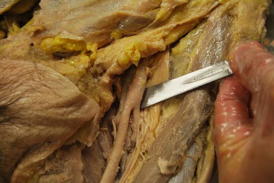
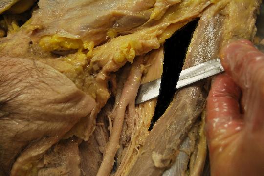
If there is a sweet spot for the Fascia Iliaca block, it is ‘The Triangle”. That’s what I have come to call it, anyway. This is not the ‘Femoral Triangle’ you were taught in medical school [nonmember]…
REGISTER for FREE to become a SUBSCRIBER or LOGIN HERE to see the full article!
[/nonmember]
[wlm_ismember]
, but it is a portion of that more famous ‘Femoral Triangle’. The traditional landmark technique (going a centimeter below the intersection of the middle and lateral thirds of the line between the ASIS and pubic tubercle) is supposed to put you here, but ultrasound is much more exacting. Let me elaborate further on this newly defined space.
I have demonstrated in a video in the Fascia Iliaca section of this website how you can go from the transverse plane of a Femoral Nerve block to the appropriate positioning for a Fascia Iliaca nerve block by turning to a longitudinal orientation of the ultrasound probe and moving in a cranial direction. There was mention of the Sartorius Muscle that could get in the way. The Sartorius Muscle is an important marker, but it can be a hindrance with the traditional and ultrasound approach.
The key to identifying the appropriate Fascia Iliaca plane by ‘pop’ or viewing local anesthetic spread is to remain in ‘The Triangle’, the lateral border of which is just medial to the medial border of the Sartorius Muscle. The medial border of ‘The Triangle’ is the femoral nerve, and the upper aspect is the inguinal ligament. The distal ‘tip’ is where the femoral nerve approaches and passed below the Sartorius Muscle. Basically, this space is the ‘exposed’ anterior surface of the Iliopsoas muscle. I will go into more detail in next week’s Tip of the Week, but notice how ‘The Triangle’ gets wider as the Sartorius Muscle moves laterally when you move cranially with the ultrasound probe in a transverse plane. You will notice this as you are lining up for a Femoral Nerve block when you are too caudad, and the Sartorius Muscle encroaches or begins to cover the Femoral Nerve.
 As you move the probe cranially from the transverse Femoral Nerve block position, you end up with more of the Iliopsoas Muscle exposed. Turn the probe 90 degrees as you watch the marbled appearance of this muscle change to multiple long and straight lines going across the screen. If you stay medial to the Sartorius Muscle, the Fascia Iliaca immediately overlying this muscle will become apparent. You should slide the probe in this orientation until you are over the ilium as described in the section of this website previously mentioned.
If you move too far medially, you will see the increased striations of the Femoral nerve. More medially, and you will see the pulsating Femoral Artery then the compressible Femoral Vein. Too far laterally, and a new muscle belly will overlie the Iliopsoas, the Sartorius. This transposition can be subtle and can easily be overlooked. If you think you’ve gone too far laterally, you can quickly turn back to the transverse plane to see the now large oval-shaped Sartorius in the way and reposition medially once more and rotate back. Another trick is to notice as you move the longitudinally oriented probe medially and laterally very slowly, you will see a white band moving across the screen back and forth. You are catching the fascial sheath around the Sartorius obliquely on the medial side of the muscle and moving it up and down the edge of the probe and, therefore, across the screen.
[/wlm_ismember]
Keep this relationship in your mind as you perform this block, and watch for the subtle signs that you are remaining within ‘The Triangle.’

