

Identifying the Subgluteal Sciatic Nerve
Let’s briefly go over how you should go about finding the sciatic nerve at the subgluteal position. First, patient positioning is important (so is yours, but ergonomics is a subject for another day). Having the patient pull their knee forward and flex at the hip will ‘tighten’ the tissue layers which will bring structures closer to the surface. Second, after identifying the greater trochanter (GT) and ischial tuberosity (IT), allow your curved probe to sit between these two structures, and allow the edges of it to rock between them. The farther down the back of the thigh that your probe scans, the more complex the image becomes as you will begin to see where the three hamstring muscles come into the picture beyond the tip of the IT. If you set your probe too high between these bony structures, the picture will become more complex as well as you will begin to see the hip abductors (superior & inferior gemellus, gluteus medius & minimus and piriformis) which presents a more complex overlapping. When you rock the probe back and forth, you will recognize a more complex image that comes and goes as you extend your scan in either direction. Honesty, you can identify the sciatic nerve between the GM and any muscle deep to it anywhere along its path, but it is easier to keep from getting lost when the image is simplified.
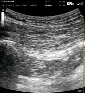
You should allow the probe to rest once you recognize this simple pattern starting superficially:
hyperechoic strip representing the skin, the long wavy lines of the subcutaneous tissue, then the striated appearance of muscle which is the gluteus maximus (GM). The bony landmarks of the greater trochanter and ischial spine will flank this image on either side. If you follow the GM deeper, you should encounter the distinct border of another muscle, the quadratus femoris (QF). Once you have this image, rock the probe until you catch the sciatic nerve at a 90 degree angle where it will best come into view. There is a good image of this on page 548 of Regional Anesthesia and Pain Medicine Nov-Dec 2013 or follow the link below to see more images. This is important since sometimes it is difficult to visualize the muscle bellies AND the sciatic nerve in the same image due to anisotropy. Of course, seeing a thick white structure that continues as you rock and slide up and down is another helpful sign…if you see this between the two most superficial muscle bellies. I have more comments on this in the Education section of the website. (see SUBGLUTEAL SCIATIC)
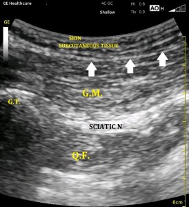
Recognize the boundary between the subcutaneous tissue and the GM (white arrows). There is a difference in the texture of these two layers, and sometimes it is helpful to see it more distinctly by moving the probe around slightly.
Remember that sometimes the sciatic nerve is not as distinct as it is in these images. Note that without identifying the boundary between the GM and QF, you may be tempted to go after one of the white areas below the QF. The sciatic nerve is usually just below the depth of the superficial aspect of the GT and IT.